
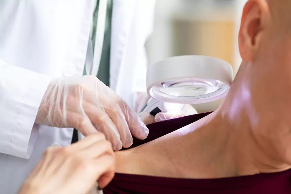
Dermoscopy is a non-invasive, diagnostic technique that allows for the magnified visualization of skin lesions, helping dermatologists accurately assess pigmented and non-pigmented skin conditions. At Ranchi Advance Skin Center, Dr. Pradeep Kumar uses state-of-the-art dermoscopy to improve diagnostic precision, especially in detecting early signs of skin cancers and other complex skin disorders.
Dermoscopy, also known as epiluminescence microscopy or skin surface microscopy, provides a clearer, more detailed look at the skin’s subsurface structures that are not visible to the naked eye. It enhances the ability to differentiate between benign (non-cancerous) and malignant (cancerous) lesions.
This advanced tool is commonly used for:
Evaluating moles and pigmented lesions
Early detection of melanoma and other skin cancers
Diagnosing conditions like psoriasis, lichen planus, scabies, and fungal infections
Assessing hair and scalp disorders (trichoscopy)
Nail disorder evaluation (onychoscopy)
Dermoscopy is quick, painless, and extremely valuable in reducing the need for unnecessary biopsies. It helps in making real-time decisions about whether a lesion needs to be removed, monitored, or left alone. When combined with clinical expertise, dermoscopy greatly increases diagnostic accuracy and patient confidence.
At Ranchi Advance Skin Center, dermoscopy is a standard part of our skin check-ups, especially for patients with multiple moles, unusual skin growths, or a family history of skin cancer. Dr. Pradeep Kumar’s training and experience ensure that every dermoscopic evaluation is thorough, evidence-based, and tailored to the patient’s needs.
Early diagnosis saves lives. Schedule a dermoscopy screening today and take the first step toward proactive skin health with trusted care.